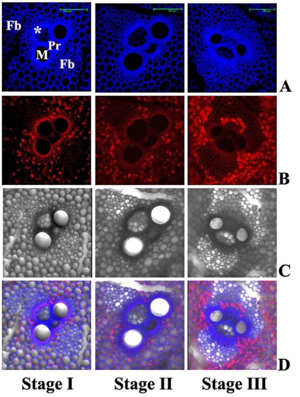
Figure 4
Download original image
CLSM images of transverse sections through internodes of B. balcooa at different stages of fiber development: Stage I. Comparatively young 2nd internodal section, fiber bundle formation initiated. Stage II. Lignification is much higher at the 4th internodal section. Stage III. Fully developed fiber sheaths at the 5th internodal section. A. Lignin and other cell wall components such as ferulic acid, a precursor of lignin, autofluoresced upon excitation at 405 nm, images acquired at 440–460–nm range. B. Chlorophyll autofluorescence in non-fibrous parenchymatous cells outside vascular and fiber bundles (excitation 458 nm, emissions at 640–700 nm). C. Same sections under bright field, showing vascular bundle with Fb (fiber bundle), M, metaxylem; Pr, protoxylem and * for phloem. D. Confocal images acquired in A (blue fluorescence) and B (red fluorescence) were overlaid on the bright field image. (A color version of this figure is available at www.afs-journal.org.)